Minimally Invasive Thoracic Surgery
- Home
- /
- Minimally Invasive Thoracic Surgery


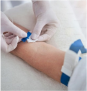
Our Department Specializes in providing Advanced Surgical Solutions for Thoracic conditions using Minimally Invasive Techniques. We are dedicated to Delivering High-Quality Care, Reducing Patient Discomfort, Minimizing Scarring, and Promoting Faster Recovery Times. Our Team of Experienced Thoracic Surgeons Utilizes State-of-the-Art Technology and Innovative Approaches to provide optimal outcomes for our patients.

Reduced Trauma and Faster Recovery

Improved Surgical Precision and Visualization

Lower Risk of Complications


Sunrise hospitals , Experience, Expertise, Care

Quality of the Care Process, Communication, follow up, staff service.
Doctors were good and they treated my mother well..
I've just had a most enjoyable experience at Sunrise Hospital.
Dr. Nassar Yusuf provided me with expert consultation, demonstrating deep expertise and care.
The doctors and nurses and everyone cares about every patient..
Everyone on nursing duty at 3rd floor was very well behaved and very caring from admission to discharge they were truly angels on earth. Special thanks to Dr Gregory Sir and everyone who helped with the surgery..

Where Compassionate Care Meets Healing Excellence.
Your Pathway to Wellness Begins Here.
Experience, Expertise, Care