About the Radiology / Radiodiagnosis And Imaging
Department

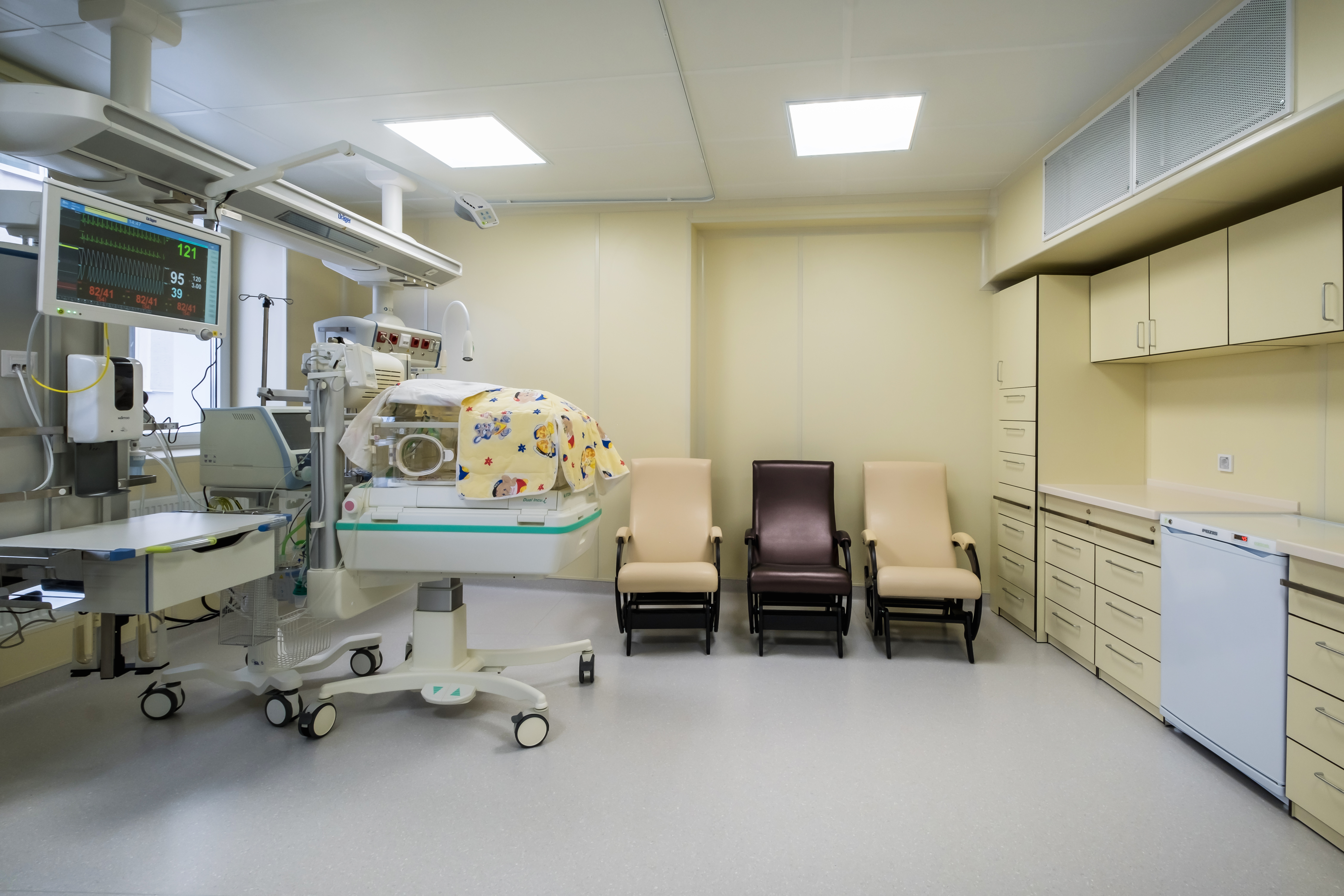
Today radiological imaging is no longer limited to the use of X-rays alone, and includes technology-intensive imaging with high frequency sound waves, magnetic fields, and radioactivity. There is also Interventional Radiology, where minimal invasive procedures are done with the guidance of imaging technologies.

The Radiology Department provides a wide range of diagnostic imaging services: including CT scanning, DEXA - Bone Density Scanning, Mammography, MRI scanning, Ultrasound scanning, and X ray.


100%

Advanced Surgical Interventions

100%

Comprehensive Disease Management

100%

Innovative Research and Education

Radiology / Radiodiagnosis And Imaging Services

Treatment and Procedure

An MRI (Magnetic Resonance Imaging) scan uses strong magnetic fields and radio waves to generate detailed images of the organs and tissues in the body. It is particularly useful for imaging soft tissues, the brain, spinal cord, and joints.

A PET (Positron Emission Tomography) scan detects metabolic signals and is often used to detect cancer and evaluate its spread. A CT scan, on the other hand, uses X-rays to create detailed images of the body's internal structures. PET-CT combines both scans for a more comprehensive evaluation.

The radiologist uses imaging techniques such as ultrasound, CT, or MRI to locate the area of concern. A needle is then inserted through the skin into the area to collect tissue samples for further examination.

Interventional radiology (IR) is a subspecialty of radiology that uses imaging techniques like X-rays, CT scans, and ultrasound to guide minimally invasive surgical procedures. Examples include angioplasty, stent placement, and biopsy.

Radiologists follow strict safety protocols, use the lowest effective dose of radiation, and employ protective measures such as lead aprons and shields. They also ensure proper patient positioning and use of advanced imaging technologies to minimize risks.

Facilities

Meet our team

Patient testimonials.

Stories of Success and Healing from Our
Minimally Invasive Surgery Patients.

FAQs

Frequently Asked Questions Answered

A CT (Computed Tomography) scan is a medical imaging technique that uses X-rays to create detailed cross-sectional images of the body. It works by rotating an X-ray source around the patient, capturing multiple images from different angles, which are then processed by a computer to generate detailed 3D images.

While both MRI (Magnetic Resonance Imaging) and CT scans are imaging techniques used to diagnose medical conditions, they differ in their principles and applications. CT scans use X-rays and are better for imaging bone and detecting acute injuries, while MRI uses magnetic fields and is superior in imaging soft tissues like the brain, muscles, and ligaments, without exposing the patient to ionizing radiation.

Bringing Smiles Back to health

Where Compassionate Care Meets Healing Excellence.

Your Pathway to Wellness Begins Here.